Wound Cultures and Biopsies
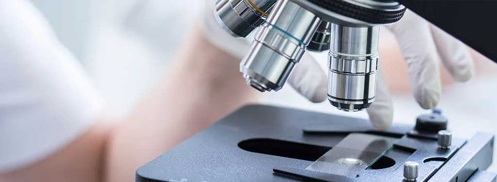
There are several available methods for obtaining a sample of a wound. A wound culture is a diagnostic laboratory test to identify microorganisms such as bacteria, fungi, or a virus that may be growing in a wound. A skin, tissue, or fluid sample is extracted from the affected area and placed in a container with a nutrient-enriched substance called growth medium or culture medium. This substance aids in the growth of organisms. If nothing substantial grows, the results of the culture are negative. Any growth of infection-causing microorganisms will yield a positive culture result. These organisms may be identified with microscopes, chemical tests, or a combination of both.
Aerobic (with oxygen) cultures are always included in wound cultures. Gram stain (direct smear evaluation) and anaerobic cultures (without oxygen) are only performed on wounds when indicated or requested by the physician. Wound cultures serve to identify and isolate the bacteria and fungi that may cause infection of the wound, as well as to identify which antibiotics will be effective in destroying any of the microorganisms identified. Cultures are performed by laboratory scientists and medical technologists who specialize in clinical microbiology.
Most wound infections contain multiple microorganisms. The most common pathogens isolated from wounds are as follows:
- Aeromonas
- Bacteroides
- Candida
- Clostridium
- Enterobacter
- Enterococci
- Escherichia coli
- Fusobacterium
- Klebsiella
- Peptostreptococcus
- Proteus
- Pseudomonas
- Streptococcus
- Staphylococcus aureus
Wound infection prevents healing, and the microorganisms can spread to other body parts as well as the blood. Septicemia (infection in the blood) can be fatal. Therefore, it is very important to identify and treat an infected wound as early as possible with an appropriate regimen of antibiotics in order to facilitate healing and prevent further complications.
The most commonly used technique is the swab culture technique, since it is the least invasive. However, it can only access organisms on the wound surface, as opposed to organisms within the tissue. Therefore, many clinicians find it to be an ineffective method of measuring infection.
Although moderately invasive and not always a feasible option, a biopsy sample is usually preferred by clinicians. For this method, the patient is given a local anesthetic prior to the removal of tissue using a cutting sheath. Bleeding is controlled throughout the process by pressure application to the wound.
Needle aspiration is a less invasive technique to use in wounds with little skin loss, such as puncture wounds. For this method, a small 22 gauge needle is inserted. In order to obtain a sample of the fluid to be biopsied, the clinician pulls back on the plunger and then changes the angle of the needle two or three times to remove fluid from different areas of the wound.
Appointment